The term 'osteotomy' refers to the surgical cutting of a bone - a 'controlled fracture'. High tibial osteotomy or HTO as it is called is a type of surgery where a cut is made towards the top of the shinbone (the tibia) with the specific objective to slightly shift the axis of weight bearing. The procedure realigns weight distribution through the knee joint away from the medial compartment of the knee affected by arthritis to the healthy lateral compartment, thus relieving pain.
High tibial osteotomy surgery is generally recommended in younger patients (under 55) affected by single (medial only) osteoarthritis of the knee. It is a Joint Preserving surgery that preserves the existing knee structures as opposed to total knee replacement surgery; and slows the progression of arthritis in the knee. This allows the young active individual to continue with his or her usual work, activities and hobbies without the limitations imposed by a Knee replacement surgery.
Should the arthritis deteriorate, a total knee replacement may still be required at a future date.
Dr Reddy may also advise a HTO in patients who have sustained a ligament injury (ACL injury or Posterolateral corner injury) as a stand alone procedure or more commonly in association with the Ligament reconstruction procedure. In patients with malalignment of the knees, this allows for a superior and more predictable outcome. HTO is also performed in conjunction with meniscal repair surgery or cartilage restorative procedures where mal-alignment of the limb co-exists.
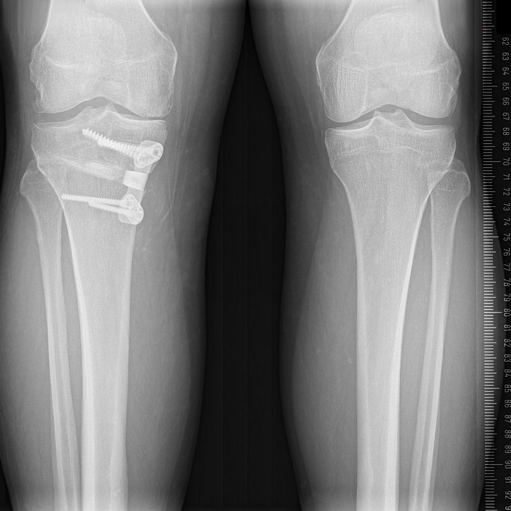
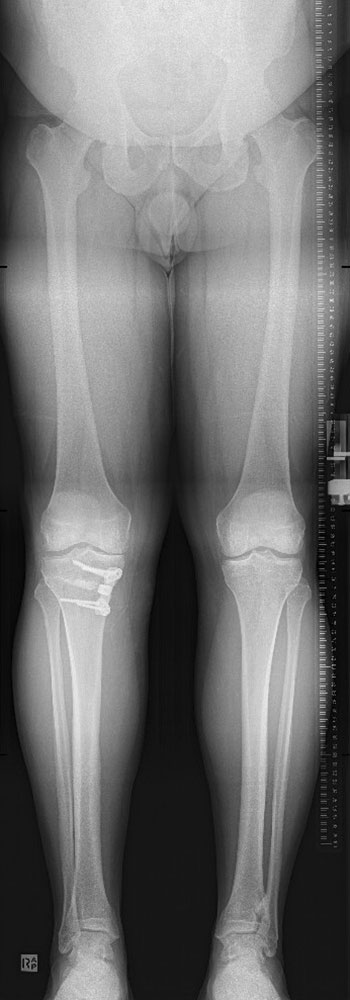
X-ray images of high tibial osteotomy fixed with plate and screws. HTO allows limb realignment to shift the weight-bearing axis, relieve pain and slow progression in early to moderate medial compartment osteoarthritis in young active individuals.
Procedure
A high tibial osteotomy can be performed either under general anaesthetic or under a spinal anaesthetic, where the patient remains awake during the procedure. The patient lies on his or her back and blood flow to the leg being operated on is restricted with a tourniquet.
Before the main procedure begins, a knee arthroscopy is generally conducted to assess the cartilage, meniscal and ligament structures and to remove any loose cartilage sections.
A 6-7cm incision to the shin is made, a precise osteotomy cut to realign the knee is made and fixed with a plate. Bone graft substitute or artificial bone graft is used to fill the gap created. Sometimes bone graft from your iliac crest may be used to enhance osteotomy healing.
High tibia osteotomy surgery generally takes around 60-90 minutes, with the patient generally able to leave the hospital within 24-48 hours.
High Tibial osteotomy (Medial opening wedge) – Things you need to know.
- Following surgery, only touch weight bearing (No weight passing through the limb) is allowed for 6 weeks.
- A hinged knee brace is worn for 4 weeks. Range of movement (ROM) is locked 0 – 60 degrees only for 2 weeks and limited to 0-90 degrees only till 6 weeks. Brace can be removed for showering and twice daily for exercises as advised by Dr Reddy and your therapist.
- You may return to sedentary/desk jobs at 3 weeks, but manual/heavy work cannot be performed for 12 weeks. Return to sports would take 6-10 months.
- Dr Reddy does not recommend or perform medial opening wedge HTO in smokers who are not willing to quit as this results in a higher than acceptable risk of non-union or the osteotomy site not healing. Obesity, involvement of other knee compartments are other contraindications to the procedure.
- Overview of risks: Risks of knee surgery include nerve injury leading to long term numbness around the knee, bleeding, infection, blood clots (DVT, PE) in the legs or/and lungs. Specific risks of HTO include non-union, persistent pain in the front of knee, joint stiffness, problems with plate and screws used to fix the osteotomy.